Transcript:
Hello. This is Dr Shaun Lehman wanting to talk today about lateral hip pain. There is a structure called the trochanteric tendon that attaches to the lateral trochan bone on the lateral portion of the hip, and here’s the top of the tendon here. The tendon structure is many different muscles that come together to form different parts of the tendons.
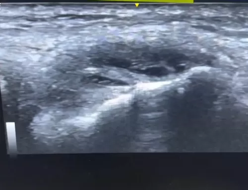
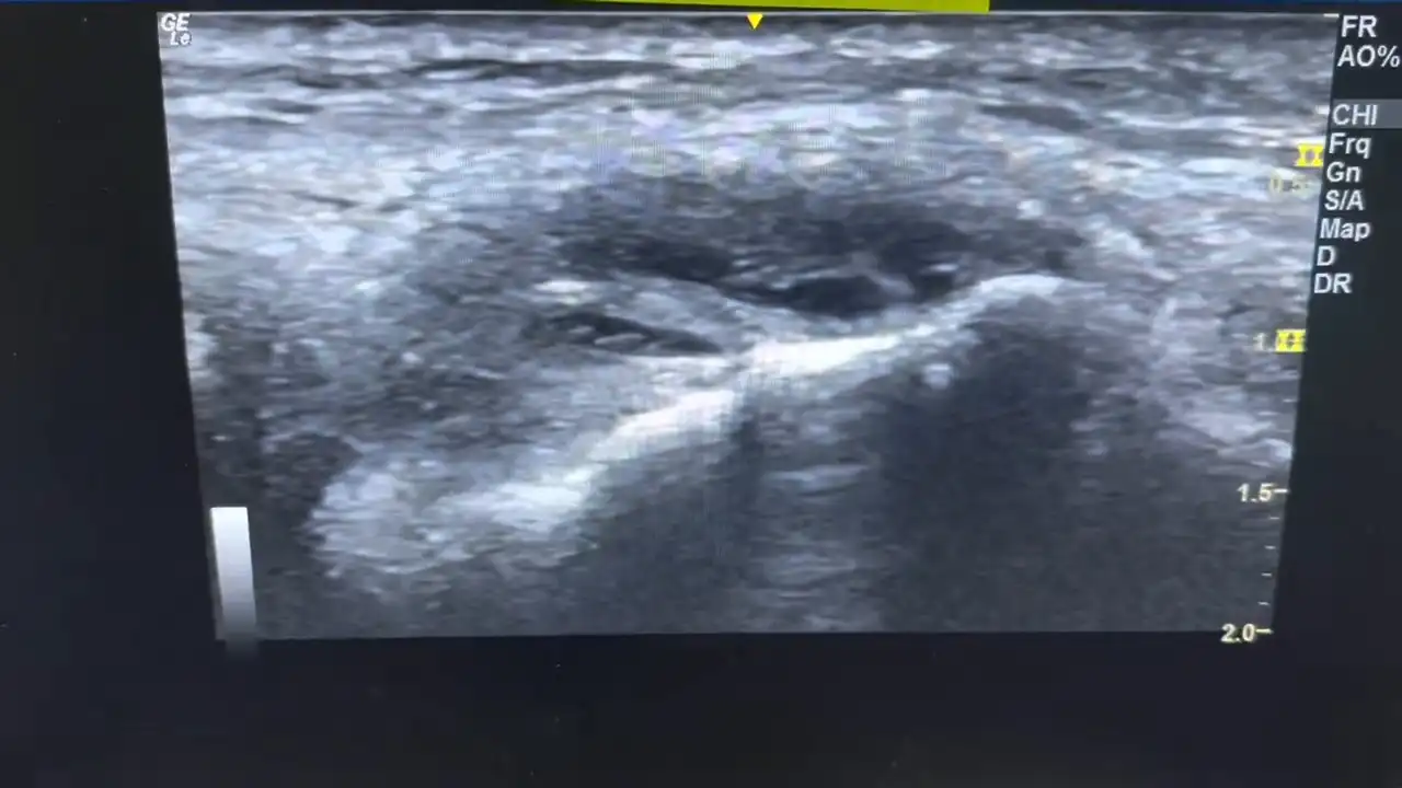
Here is a portion of dark area or hypoechoic area that may have an issue with integrity for attachment. And there here’s a view of some possible linear tears as well. And back where we started, This area right here is very interesting. There is a large tear here in the deeper portion of the muscles that attach, and it is also measured here at three point two five centimeters.
So it’s a substantial tear. This could cause, instability in the hip with one legged standing. What structure could this be? As you can see, as you come down lateral and inferior, the deeper structures are these muscle muscles right here. This most likely is the quadratus femoralis, which, is quite a wide muscle that’s deep.
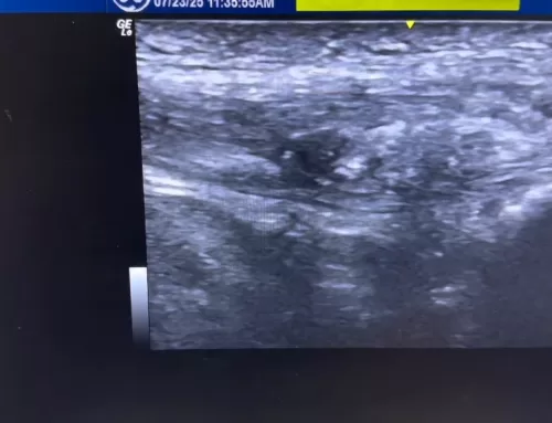
It also could be the obturator internae or gemelli, but most likely, it’s the quadratus femoralis that’s that’s tor.
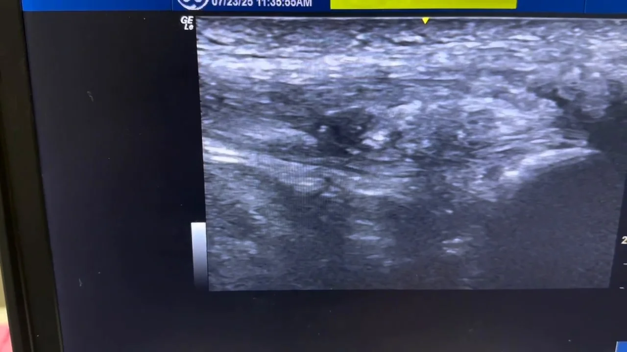
So as you can see here, the external muscles that attach here, glute maximus, and they don’t show the glute minimus, which attaches up here, and glute med, which attaches up here. Minimus is underneath, and the medius is kinda between between the two. And they do attach over here on the troch as well. So as you can see, a lot of muscles attach here.
So it’s a very busy area and, very interesting case, and I just wanted to just show you this. Of course, there are some different treatments for this, including PRP, prolotherapy. It’s very difficult to knit together a large tear. However, the the pain can definitely be treated with those treatments. If, a person had a very large tear, surgical options obviously would be in in in order.
Thank you.
