
Summary:
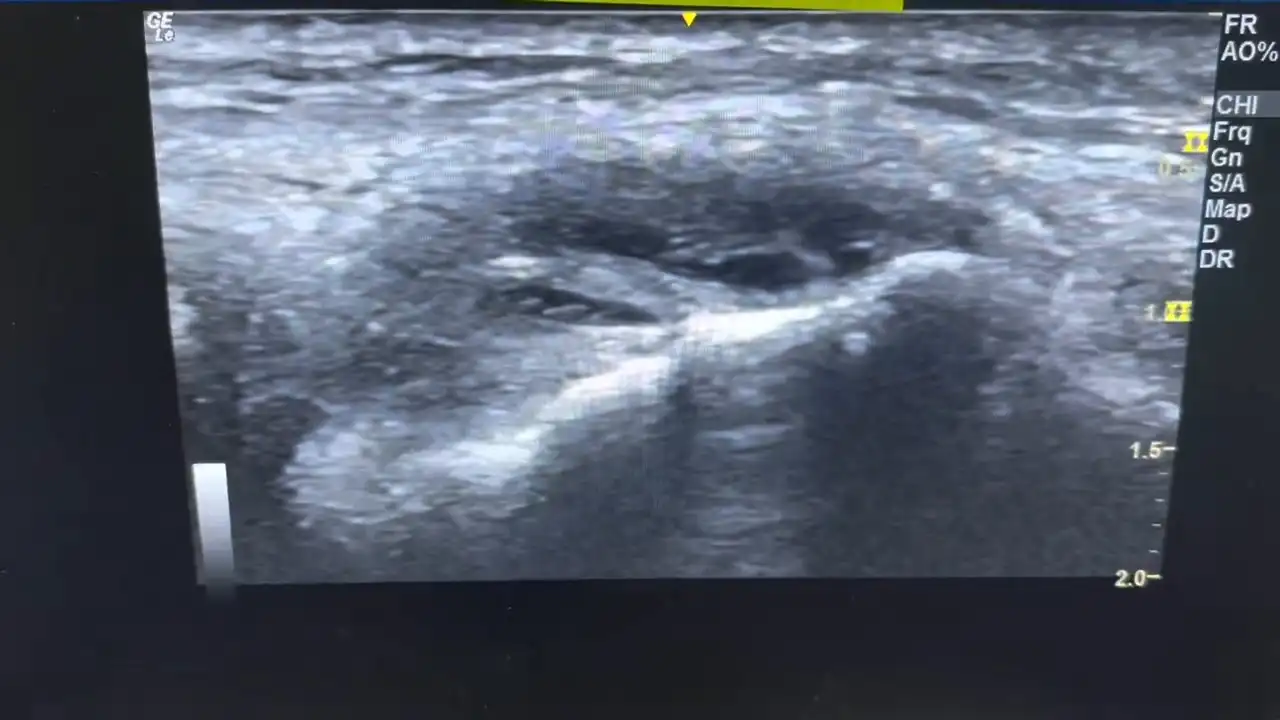
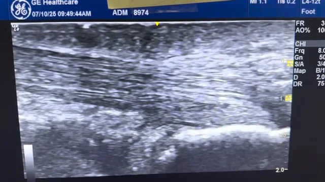
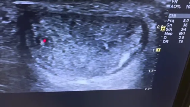
This video highlights a case of infrapatellar tendinopathy, a condition affecting the tendon just below the kneecap (patella). Ultrasound imaging reveals a swollen and irregular section of the tendon, with signs of microtears and hypoechoic (dark) areas—indicating poor healing rather than active inflammation.
Unlike traditional “tendonitis,” tendinopathy refers to a degenerative condition, often due to the tendon’s inability to fully repair itself. In this case, the most painful area aligns with visible irregularities and tissue breakdown, while other sections of the tendon appear more normal but still show minor underlying issues.
This informative video is ideal for anyone dealing with knee pain, especially athletes or individuals with jumper’s knee. It emphasizes the importance of diagnostic imaging to accurately pinpoint the source of discomfort and better understand the healing challenges involved in tendon-related injuries.