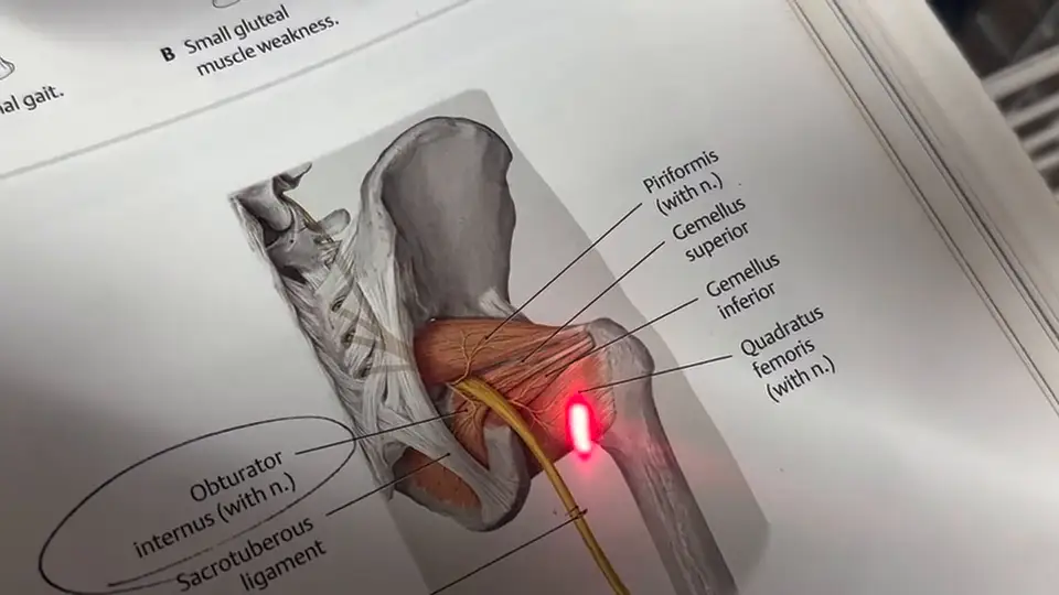
Summary:

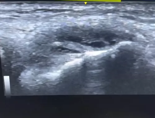
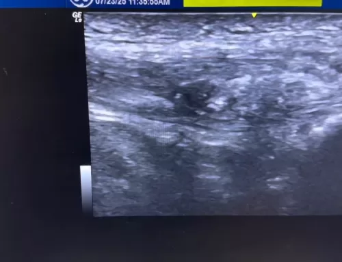
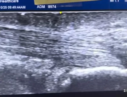
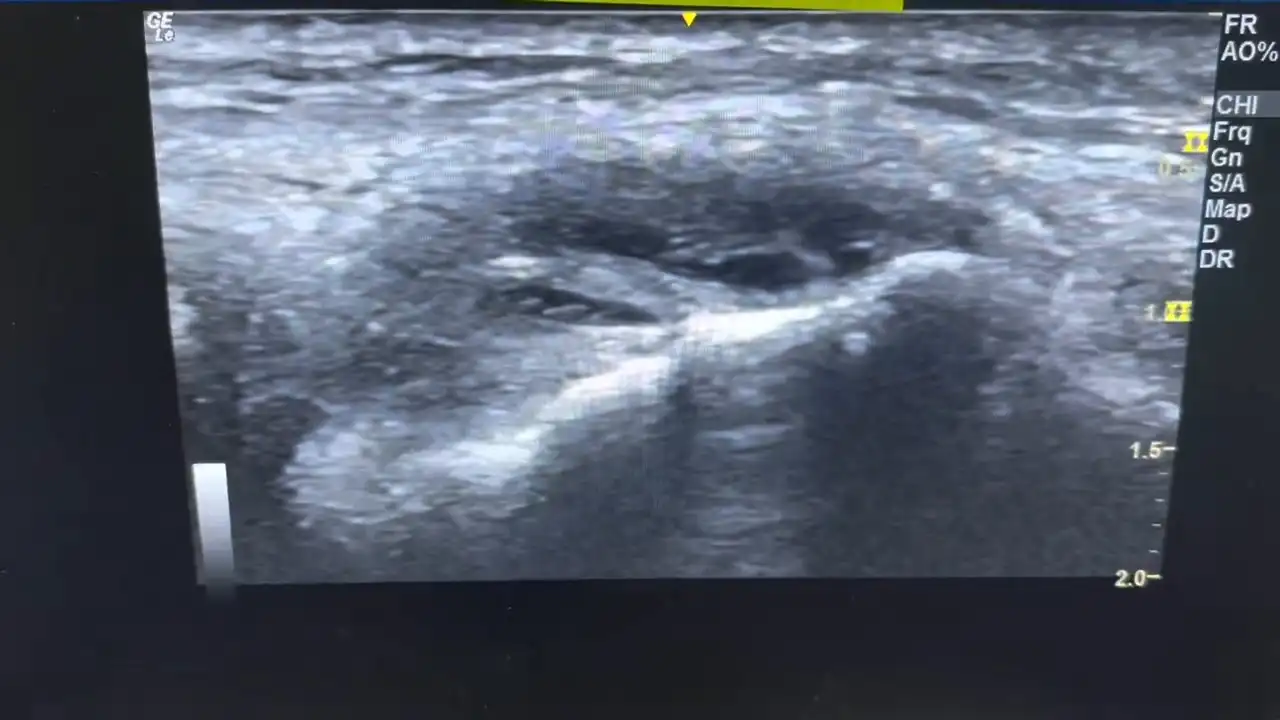
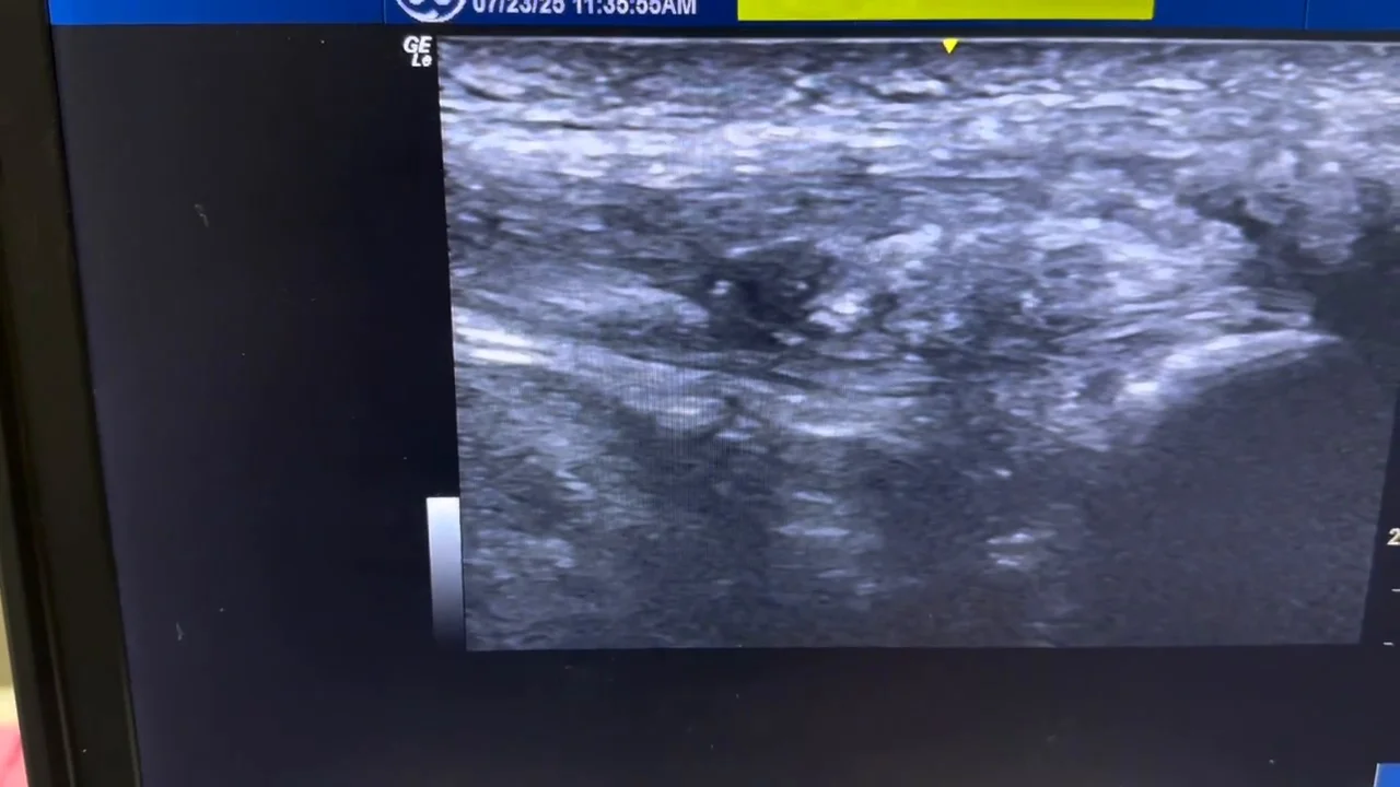
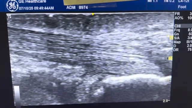
In this video, we explore Achilles tendinopathy, a condition marked by the thickening and irregular structure of the Achilles tendon due to poor healing. Using ultrasound imaging, the video highlights the distinct features of a tendinopathic tendon, including irregular fibers, hypoechoic (dark) spots, calcification, and a noticeably thicker cross-section—up to 1.38 cm, compared to a normal, pain-free Achilles tendon which measures only 0.51 cm.

Viewers can clearly see the contrast between healthy and damaged tissue, with the tendinopathy tendon appearing swollen, disorganized, and “fish belly”-like in shape. This side-by-side comparison is valuable for anyone experiencing Achilles pain, healthcare professionals, or those interested in tendon health and sports injury recovery. Watch the full breakdown to better understand what Achilles tendinopathy looks like and why it causes persistent pain.


Transcript:

Hi, this is Dr. Sean Lehman, and today, we’re gonna talk about Achilles tendinopathy, a true tendinopathy. And what you see is a thickening of the Achilles tendon.

For some reason, the tendon seems to have a problem healing correctly, and so adaptation is growing thicker.

So at the end, we’ll show you the normal size. But you can see the size between here and here is very thick and almost looks like a fish belly or like a minnow. The fibers here are irregular. So the fibers are going left and right.

You can kind of see that dip down right here and some irregularity in it. So that’s the thickening. That’s the area that’s going be extremely painful.

Another view of that – Cross section, some hypoechoic dark spots there. You can see this part again, cross section. This dark spot is kind of weakened part of some of the tissue there. Some irregularity. This is the circle.

This is the Achilles. And some irregularity you see there.

Let me go through these quick as well. There’s two little calcification and little dark spots there as well.

And then this is the thickening where you can kind of see that little calcification area.

We did measure it. That’s one point three eight centimeters. You can see down here. So this is we’re going to compare to the normal side.

This doesn’t hurt.

And there you go. That’s like zero point five one centimeters. So you can see that how the fibers go left to right. That’s the top. Here’s the bottom.

And the fibers look nice and uniform. They’re not thick.

So here’s a great example of normal Achilles versus the tendinopathy Achilles that was super thick and irregular. Thank you.