
Summary:
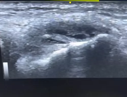
In this video, we examine a unique case of Achilles pain that doesn’t stem from typical tendon thickening, but instead from extra bone growth at the heel—right where the Achilles tendon attaches. At first glance, the tendon appears healthy and not thickened, misleading both patients and providers.
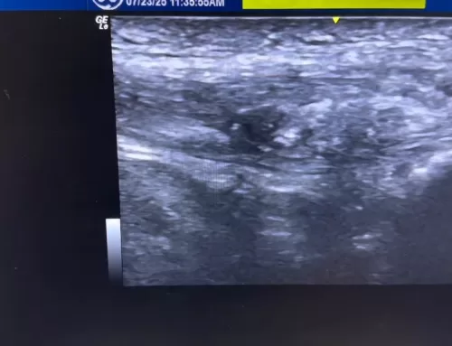
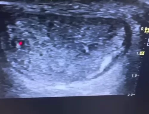
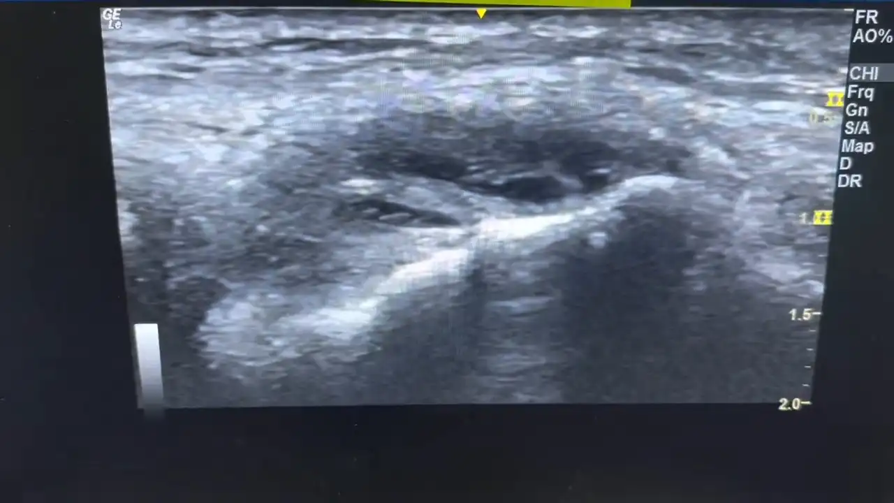
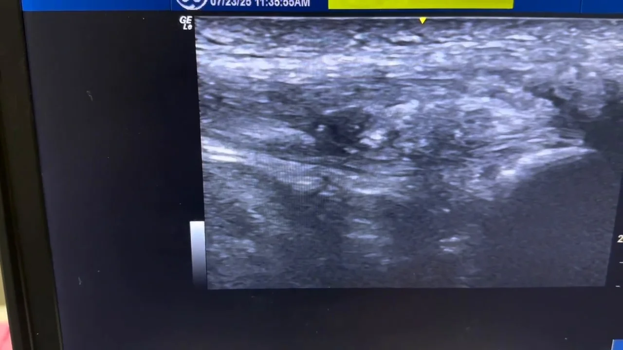
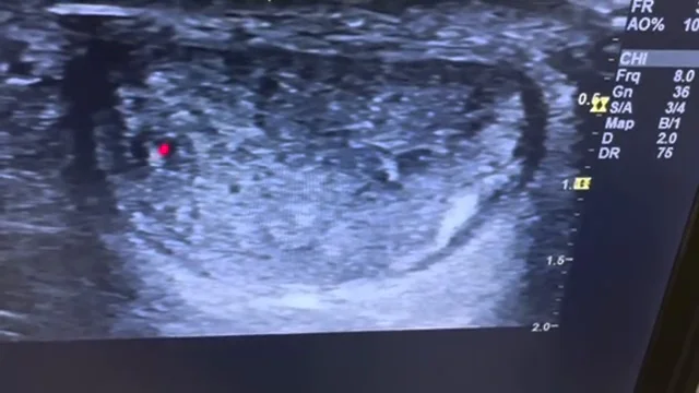
However, deeper ultrasound imaging reveals a prominent bony bump, known as insertional Achilles tendinopathy, which is the true source of the patient’s pain and swelling. The affected heel shows irregular bone structure, hypoechoic (dark) areas, and localized inflammation right at the pain point.
The comparison with the other (non-painful) side shows only minor tendon changes, confirming that the extra bone and tendon irregularity at the insertion point is the root cause of discomfort. This video is a great resource for understanding less obvious causes of heel pain and how ultrasound imaging can reveal hidden structural issues often missed in a surface-level exam.