Summary:
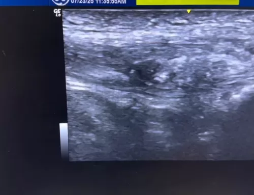
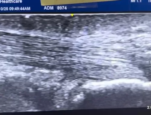
In this video, we explore Achilles tendinopathy, a condition marked by the thickening and irregular structure of the Achilles tendon due to poor healing. Using ultrasound imaging, the video highlights the distinct features of a tendinopathic tendon, including irregular fibers, hypoechoic (dark) spots, calcification, and a noticeably thicker cross-section—up to 1.38 cm, compared to a normal, pain-free Achilles tendon which measures only 0.51 cm.
Viewers can clearly see the contrast between healthy and damaged tissue, with the tendinopathy tendon appearing swollen, disorganized, and “fish belly”-like in shape. This side-by-side comparison is valuable for anyone experiencing Achilles pain, healthcare professionals, or those interested in tendon health and sports injury recovery. Watch the full breakdown to better understand what Achilles tendinopathy looks like and why it causes persistent pain.


